

- Description
- Reviews (9)
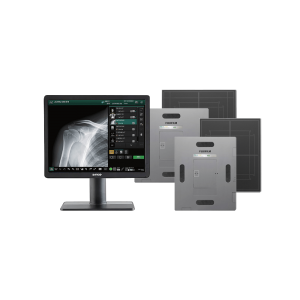
Description
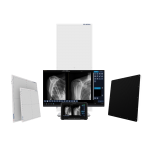
DELWORKS EDR
DELWORKS
Pre-exposure display of patient and procedure information, x-ray generator exposure factors, status and control functions integrated into a single display screen
Positioning guide: queues proper positioning for each view
Deviation Index (DI): standard reporting to assist in dose minimization
Exam-specific image processing algorithms automatically optimize images based on selected patient anatomy
Enhanced image processing parameters: APR specific default values and manual adjustment, if desired
Image rotation in 90° steps, horizontal mirroring, automatic and manual image cropping to collimated area
Full array of measurement tools
Easy verification and correction of image laterality and patient orientation
Intuitively add orientation markers and text comments directly to acquired images (pre-defined or free text)
PICC tool applies enhanced processing to images for better visualization of leads, lines, or tubes
Effective management, analysis, and reporting of rejected images
Detailed histograms of pixel density
User selection of modified LUT (Look-Up Table) based on various exam types
Manual adjustment of the LUT, window, and level
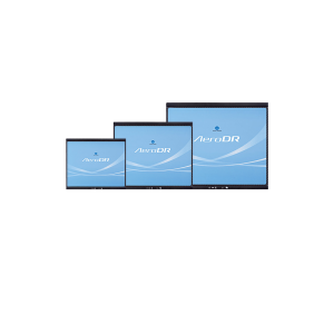
Premium Options Available in Single, Dual, or Multi-Detector Applications
E14C
Ultra-light, portable 14 x 17 in (35 x 43 cm) detector with outstanding image quality and offer the compact versatility needed to optimize workflow.

E24C
Ultra-light, portable 10 x 12 in (24 x 30 cm) Cesium wireless detector with outstanding image quality that offers the compact versatility needed to optimize workflow.

E17C
A large-format 17 x 17 in (43 x 43 cm) fixed detector designed to efficiently minimize technologist interaction with upright exams in both a dual or a multi-detector configuration

Monolithic long length 17 x 42 in (43 x 107 cm) detector for full spine and long leg imaging eliminates potential stitching misalignments, improves workflow, and decreases the patient dose

EasyConnect
DELWORKS E-Series Wireless Detectors feature EasyConnect — an Auto Exposure Detection (AED) technology that keeps the detector in a standby mode, awaiting exposure from any X-ray source. Once an exposure is detected it instantly captures the x-ray image and transmits it wirelessly to the system workstation

The DELWORKS FIT Portable Tablet Workstation aims to maximize the portability and efficiency of the DELWORKS software — utilizing it anytime, anywhere.
The user-friendly features of the DELWORKS software are beautifully translated to a rugged tablet PC, capable of withstanding even the harshest radiology (or non-radiology) environments. This handheld workstation features wireless connectivity, plenty of robust, secure solid-state storage space, and a powerful Intel® Core™ i5 processor to perform rapid and productive patient side studies, without limitations.
The DELWORKS Laptop is an option for those who prefer portability combined with built-in keyboard input. This system includes a robust 500 GB Solid State Drive (SSD) and a full HD 15.6” display for secure storage and outstanding image display quality.


Quality Assurance
The DR QC Phantom allows comprehensive quality assurance testing for DR Detectors and associated software. The phantom enables seven quality assurance assessments for monthly and semi-annual evaluation, including exposure linearity and sensitivity, high and low contrast reproducibility, artifact and residual image detection, image resolution, collimator beam alignment, measurement tool accuracy, and display jitter QA testing.
TECHNICAL SPECIFICATIONS
Detectors
| DETECTORS | E14CW | E24CW | E17C | LLI |
|---|---|---|---|---|
| DETECTOR SIZE | 14 x 17 in (35 x 43 cm) | 24 x 30 cm | 17 x 17 in (43 x 43 cm) | 17 x 42 in (43 x 107 cm) |
| SCINTILLATOR | Cesium Iodide (CsI) | Cesium Iodide (CsI) | Cesium Iodide (CsI) | Gadolinium Oxysulfide |
| ACTIVE AREA | 13.8 x 16.8 in | 8.7 x 11.2 in | 17 x 17 in | 17 x 42 in |
| PIXEL MATRIX | 2400 x 2880 | 1646 x 2057 | 2860 x 2874 | 3072 x 7680 |
| PIXEL PITCH | 148 μm | 148 μm | 148 μm | 140 μm |
| DIMENSIONS | 15.1 x 18.1 0.6 in | 10.6 x 13 x 0.6 in | 19.7 x 19.3 x 1.7 in | 18 x 44 x 0.8 in |
| WEIGHT | 6.2 lbs w/battery | 3.5 lbs w/battery | 27.5 lbs | 24 lbs |
| BIT DEPTH (A/D) | 16 bit A/D | 16 bit A/D | 16 bit A/D | 16 bit A/D |
| IMAGE DISPLAY | 3 seconds | 1 second | 4 seconds | 9 seconds |
| CYCLE TIME | 9 seconds | 6 seconds | 6 seconds | 12 seconds |
| EXPOSURE WINDOW | Up to 3.2 seconds | Up to 3.2 seconds | Up to 3.2 seconds | |
| AUTO EXPOSURE DETECTION | Yes | Yes | No | Yes |
| WIFI | 802.11 a/b/g/n 2.4 GHz / 5 GHz |
802.11 a/b/g/n 2.4 GHz / 5 GHz |
N/A | 802.11 a/b/g/n 2.4 GHz / 5 GHz |
| DROP SENSOR(S) | Yes | Yes | No | |
| BATTERY TYPE | Li-Ion 3.35 Ah 7.4 V | Li-Ion 3.35 Ah 7.4 V | N/A | Li-Ion |
| LIMITING RESOLUTION | 3.5 lp/mm | 3.5 lp/mm | 3.5 lp/mm | |
| DQE | 70% @ 0 lp/mm 51% @ 1 lp/mm 39% @ 2 lp/mm 23% @ 3 lp/mm |
66% @ 0 lp/mm 51% @ 1 lp/mm 39% @ 2 lp/mm 23% @ 3 lp/mm |
66% @ 0.5 lp/mm 52% @ 1 lp/mm 42% @ 2 lp/mm 25% @ 3 lp/mm |
60% @ 0 lp/mm 45% @ 1 lp/mm 32% @ 2 lp/mm 15% @ 3 lp/mm |
| MTF | 61% @ 1 lp/mm 31% @ 2 lp/mm 15% @ 3 lp/mm |
61% @ 1 lp/mm 31% @ 2 lp/mm 15% @ 3 lp/mm |
64% @ 1 lp/mm 32% @ 2 lp/mm 17% @ 3 lp/mm |
65% @ 1 lp/mm 33% @ 2 lp/mm 18% @ 3 lp/mm |
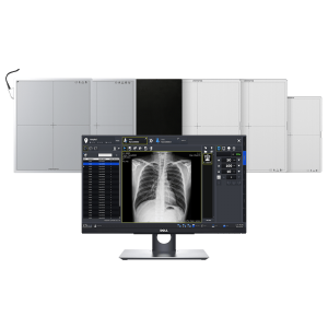
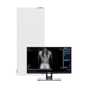
Desktop Workstation
Desktop Workstation (Monitor)
Tablet Workstation
Laptop Workstation

Reviews
There are no reviews yet.